
a handheld dental X-ray machine is a tube, a battery — and a sensor
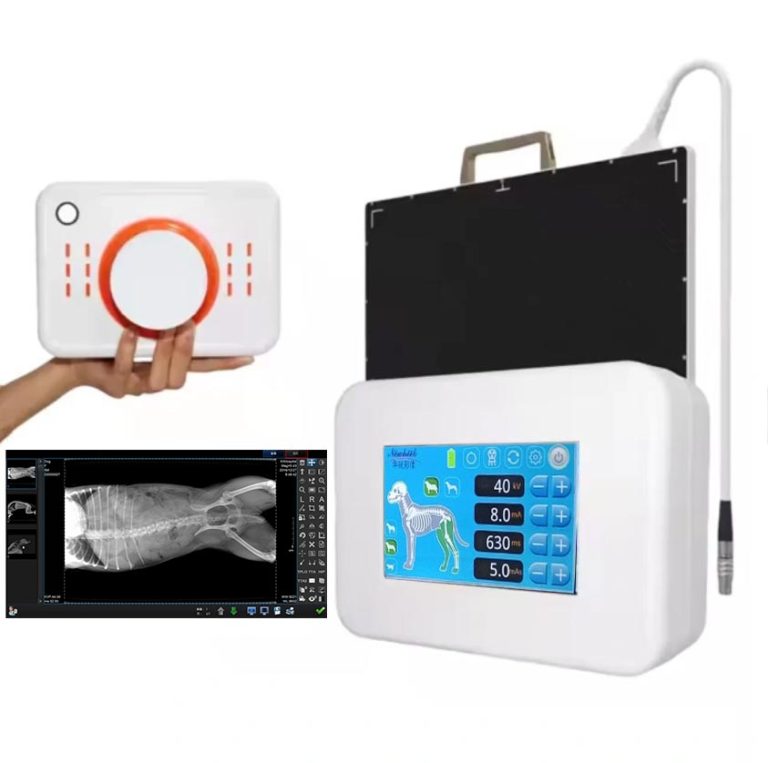
A handheld dental X-ray machine is an operator-held intraoral X-ray source: a compact tube head on a rechargeable battery grip, with a beam-limiting cone and an integrated backscatter shield. But the part that decides whether the image is diagnostic is the receptor it exposes — an intraoral sensor that is, in engineering terms, a small flat panel detector. So a buyer’s guide for handheld dental units is really a guide to two things: the X-ray source you hold, and the detector that turns the beam into a picture. This article covers both, with the detector side told the way a flat-panel specialist would.

What the unit actually is
A handheld dental X-ray machine has four defining parts:
- Tube head with beam-limiting cone (PID). A fixed or selectable voltage — commonly in the 60–70 kVp range across the category — collimated to a small field over the receptor.
- Rechargeable battery grip. The handset houses a lithium-ion pack sized for a full clinical day; a control panel sets exposure time.
- Backscatter shield. A clear lead-equivalent shield at the cone edge that blocks radiation scattered back from the patient. On compliant units it is not removable.
- Exposure trigger and indicators. A trigger with audible and visual status confirmation, so the operator always knows when radiation is being produced.
The fifth, invisible part is the receptor — and that is where this article’s expertise lives.
Why the detector is the variable that matters
For intraoral views — periapicals and bitewings — the handheld unit and a wall-mounted unit expose the same class of sensor at similar kVp and mA. The difference a buyer feels is almost never the source; it is the receptor. Two detector facts dominate the outcome:
- Resolution. Intraoral sensors in the category typically resolve about 20–33 line pairs per millimetre (CMOS). Finer resolution means sharper endodontic file measurements and clearer periapical detail.
- Active area. Size 1 (about 24×40 mm) suits anterior and paediatric work; Size 2 (about 31×41 mm) covers most adult periapical and bitewing needs. A sensor that is the wrong size for your patient mix forces retakes — and retakes are the real dose cost.
Our primer on what detector specification numbers mean in the exam room explains the rest of the datasheet — pixel pitch, bit depth, scintillator — in the same plain language.
CMOS versus CCD, and why backplane choice shows up here
Most modern intraoral sensors are CMOS, and the reason is the same one that drives larger panels: a CMOS backplane reads the pixel array faster and with less noise than the older CCD approach. If you want the deeper technical picture, our article on how flat panel detector backplanes differ covers a-Si, IGZO and CMOS side by side. For a handheld workflow the practical takeaway is simple: a CMOS intraoral sensor delivers a usable image quickly, which is exactly what chairside dentistry needs.
Wired or wireless sensor?
The same trade-off that shapes general radiography applies at the intraoral scale. A wired sensor has no battery to charge and no wireless link to fail; a wireless sensor removes the cable snag around a patient’s mouth and a moving operator. For a handheld unit — where the operator is already holding the source — wireless is often the better ergonomic match, but only if the wireless link and battery autonomy are documented, not assumed. Our wired vs wireless comparison lays out the decision variables.

Dose: the ALARA relationship you cannot skip
A handheld unit is an ionizing-radiation source, and its safety case is identical in principle to a wall-mounted system’s: keep every exposure as low as reasonably achievable (ALARA). The published numbers that matter:
- Backscatter shield. Guidance summarized from the NCRP’s dental radiology report requires an FDA-cleared handheld unit to carry a backscatter shield of at least 0.25 mm lead equivalence, kept clear and non-removable.
- Operator dose is very low. Studies evaluating handheld devices report average operator exposure of about 0.045 mSv per year — under 1% of the 50 mSv annual occupational limit set by the NCRP (figures as summarized in Dimensions of Dental Hygiene from NCRP Report No. 177 and related studies).
- Rectangular collimation cuts patient dose. Where used, patient-absorbed dose drops by roughly 50%, per the same NCRP-based guidance.
- Leakage is capped. FDA guidance for hand-held X-ray equipment and IEC 60601-1-3 set leakage-radiation limits (0.88 and 0.25 mGy per hour respectively) that compliant units must meet.
Technique still decides real-world dose. Correct positioning — arms extended, the unit held parallel to the patient and perpendicular to the floor, backscatter shield seated close to the patient, hands never in the primary beam — is what converts good engineering into low exposure. Arm fatigue over repeated exposures degrades positioning, so training is not optional.
Handheld vs. wall-mounted: same sensors, different room
| Dimension | Handheld unit | Wall-mounted unit |
|---|---|---|
| Installation | None — charge and go | Wall mounting, arm fitting, calibration |
| Intraoral image quality | Comparable — same sensor class | Comparable |
| Best fit | Chairside, multi-room, mobile and outreach care | Dedicated radiography rooms, high-volume work |
| Panoramic / CBCT | Not possible | Available on dedicated extraoral systems |
| Throughput ceiling | Battery and handling constrain very high volume | Always in position; no battery cycle |
Compatibility you must verify before buying
A handheld unit is only as useful as the receptor and software around it. Before purchase:
- Confirm DICOM and TWAIN driver support for your imaging platform — and test it, do not assume it. Our explainer on whether a flat panel detector contains a DICOM system is the right background.
- Confirm the sensor works with the unit’s exposure range — adult and pediatric, anterior and posterior — not just with the demo sensor.
- Confirm practice-management integration. The image has to land in the patient record at the chair. Our guide to installing and using dental sensors covers the workflow details.
Specifications decoded (typical category ranges)
Ranges below are typical across the category as published in independent buyer’s guides. They are not promises about any one product — always verify against the manufacturer’s specification sheet.
| Parameter | Typical range | Why it matters |
|---|---|---|
| Tube voltage | 60–70 kVp | Higher kVp penetrates dense posterior structures |
| Tube current | 2–3 mA | Paired with kVp to set exposure and dose |
| Exposure time | 0.01–0.5 s | Shorter exposures reduce motion blur and retakes |
| Focal spot | ~0.4 mm common | Smaller spot sharpens endodontic detail |
| Sensor resolution | ~20–33 lp/mm (CMOS) | Finer detail for periapical pathology and file measurement |
| Sensor active area | Size 1 (24×40 mm) / Size 2 (31×41 mm) | Size 2 covers most adult periapical and bitewing needs |
| Unit weight | ~1.5–3 kg | Held steady through repeated exposures |
| Battery endurance | ≥100 exposures per charge (typical target) | Must cover a standard clinical day |
| Dose per exposure | ~2–5 µSv (verify per unit) | Basis for patient-dose and ALARA records |

Where dental flat panel detectors fit this workflow
A handheld dental X-ray machine is a point-of-care source; the intraoral detector is the component that determines whether the picture is diagnostic. For practices specifying the receptor itself, our dental-specific resources go deeper:
- Application of dental flat panel detectors — where and how they are used across dental imaging.
- Dental digital DR detectors — the receptor class behind modern intraoral and extraoral dental work.
- How to choose a DR flat panel detector — the general selection framework.
- Durability specs that decide where a detector really works — ingress protection, drop and load, for units that travel.
- Portable X-ray imaging and using a flat panel detector in a mobile X-ray machine — the broader point-of-care context.
FAQ
Are handheld dental X-ray machines safe?
Yes — when the unit is cleared for handheld use and operated per the manufacturer’s protocol. Compliant units carry a non-removable backscatter shield of at least 0.25 mm lead equivalence, and published studies summarized under the NCRP’s dental guidance report average operator exposures of roughly 0.045 mSv per year, under 1% of the 50 mSv annual occupational limit.
Do handheld units produce the same image quality as wall-mounted systems?
For intraoral images such as periapicals and bitewings, quality is generally comparable because both expose the same sensor class at similar parameters. Handheld units cannot produce panoramic or CBCT images; those need dedicated larger equipment.
What regulations apply to handheld dental X-ray machines?
In the United States, FDA 510(k) clearance plus state radiation-control registration, with state-specific training and shielding rules. Internationally, look for IEC 60601-series conformity, including 60601-1-3 and 60601-2-65. Always confirm your own jurisdiction’s requirements before purchase.
Does the detector need to be wireless?
Not necessarily. A wired intraoral sensor has no battery or wireless link to manage; a wireless sensor removes the cable snag at the chairside. The right choice depends on your workflow — our wired vs wireless guide covers the trade-offs.
How many exposures per battery charge should I expect?
Buying guides commonly set the bar at 100+ exposures per charge for a standard clinical day, with a 2–3 hour recharge. Actual ratings vary by model — match the rated capacity to your daily image volume.
Specifying the detector for a handheld dental workflow, or matching a dental flat panel detector to your practice? Our team answers specification questions in writing across medical, veterinary and dental imaging. Email: admin@newheek.cn | WhatsApp: +86 19062611512
你好