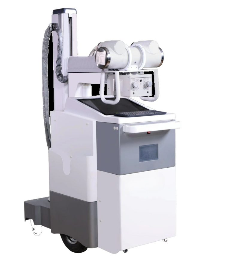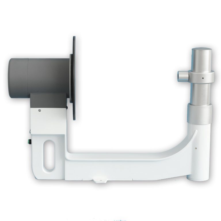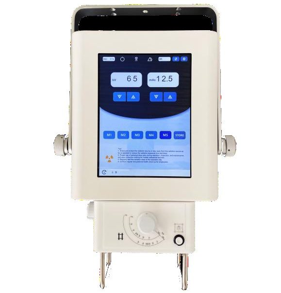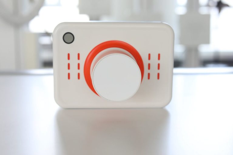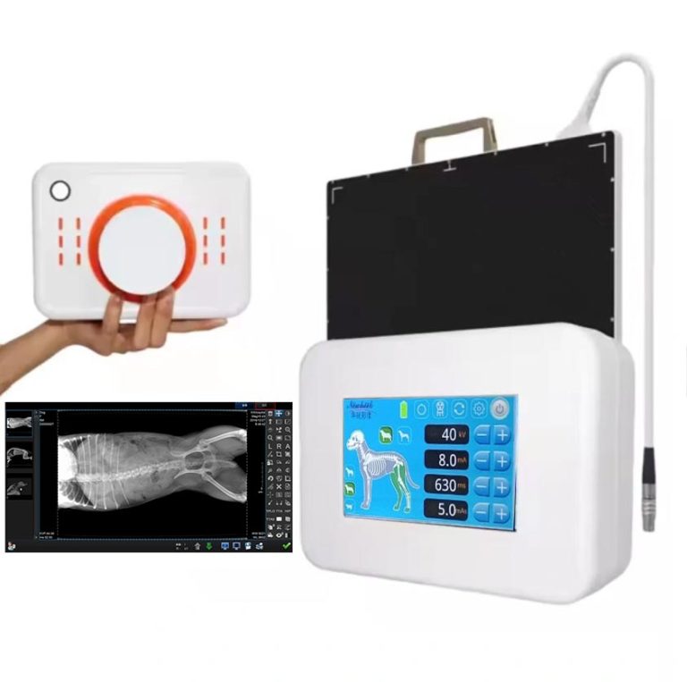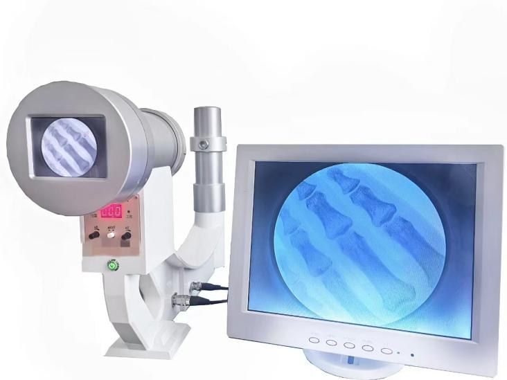
Quick answer: there is no single “veterinary X-ray machine” — there are four equipment configurations that solve four different practice problems. A portable generator in a trolley case solves the farm call; a mobile table-plus-generator set solves the multi-room clinic; a combined unit on castors solves the budget-conscious single room; a fixed veterinary DR system with a floating table solves the high-volume hospital. Choosing wrong is expensive in both directions: too little machine and your team improvises every study, too much machine and you pay for capacity that sits idle. This article walks through what each configuration is actually for, what changes when the patient is an animal rather than a person, and a five-question checklist to match configuration to caseload.
Why veterinary radiography is not “human radiography on smaller patients”
Before comparing configurations, it helps to be honest about what makes animal imaging different, because every configuration choice below traces back to one of these facts:
- The patient will not cooperate, and cannot be instructed. No breath-hold cues, no “stay still” commands that a scared cat or a painful dog will obey. Positioning is done by restraint, support, sedation, or clever equipment — usually a combination.
- Somebody is in the room holding the animal. In human imaging, staff step behind a barrier. In veterinary imaging, an owner or technician is frequently beside the table, which makes radiation protection a workflow design problem, not just a badge-and-apron problem.
- Size range is extreme. The same practice may image a guinea pig and a Great Dane in one morning. That stretches detector coverage, table capacity assumptions, and generator output requirements across a range no human clinic sees.
- Fur, dirt, and thickness vary wildly. A matted coat, a muddy leg, or a deep-chested breed changes exposure and image quality in ways a human workflow never encounters.
- Many views are taken with the animal lying down or standing on the table, so the table itself — its height, surface, and how a panel slides under or sits in a tray — matters more than in a human room where patients stand at a wall stand.
Keep those five facts in mind. Each configuration below is essentially a different answer to them.
Configuration 1: the portable generator in a trolley case
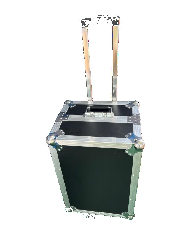
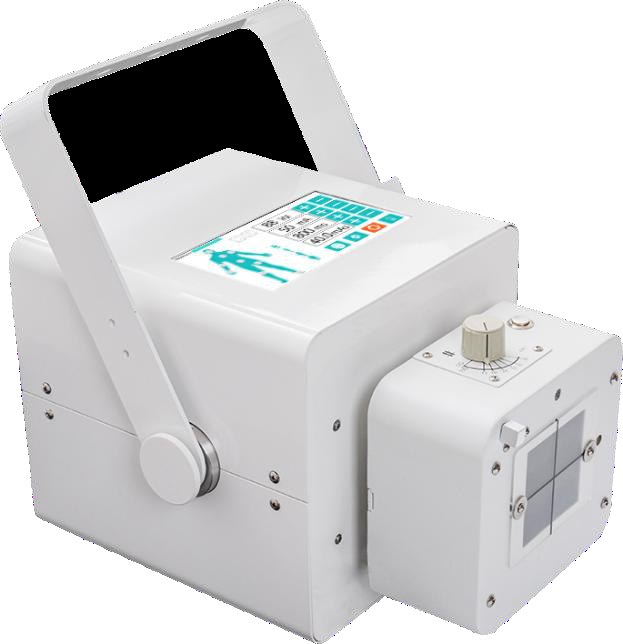
This is the smallest footprint in the veterinary line: a portable X-ray generator that packs into a wheeled case, ready to travel. It exists for the practices where imaging does not happen at a fixed table — ambulatory and equine work, farm and stable calls, zoo and wildlife handling, disaster and field screening, and mobile clinics that serve multiple locations.
The trade is deliberate: you give up the table, the restraint fixtures, and the dedicated room, and in exchange the machine goes where the animal already is. For a horse with a suspected lameness, hauling the patient to the clinic is neither practical nor safe — the generator comes to the horse, a panel goes behind the limb, and the study happens in the stable aisle.
Who it fits: ambulatory veterinarians, equine and farm-animal practices, mobile screening services, and small clinics that only occasionally need radiography and cannot justify a dedicated room. Who it does not fit: any practice doing daily, high-volume studies — packing and unpacking a case every day gets old by the second week.
For a deeper comparison of the portable class against other modalities — including where a portable fluoroscopy unit or a handheld dental unit belongs — see the site’s guide to the three different devices sold under one handheld name.
Configuration 2: the mobile table, generator and panel set
This configuration pairs a wheeled table with a portable generator and a flat panel detector, so the whole imaging setup can move between rooms instead of the patient moving to radiography. It occupies the middle ground: more capability than a bare portable, more flexibility than a fixed room.
It fits practices with multiple imaging locations inside one building — a treatment room that doubles as an imaging space, a ward side where recumbent post-op patients need studies without transport, isolation wards where moving animals across the clinic is a biosecurity problem. The reasoning is the same as in human bedside imaging: when the patient is better off not moving, the equipment moves instead. That logic is laid out in detail in the article on what a mobile X-ray machine actually does at the bedside.
Who it fits: multi-room hospitals, practices with recumbent or post-surgical patients, quarantine and isolation situations, and growing clinics that are not ready to commit permanent floor space to a radiography room.
Configuration 3: the combined unit on castors, with a cassette tray
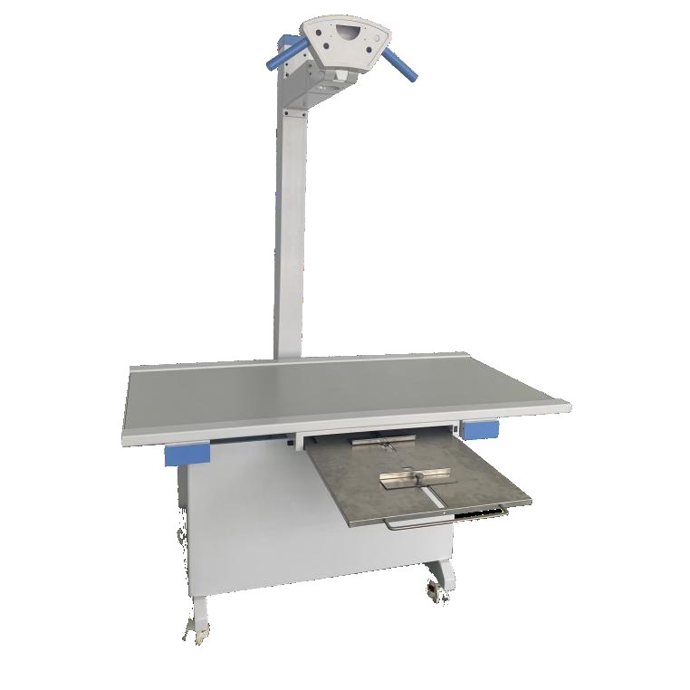
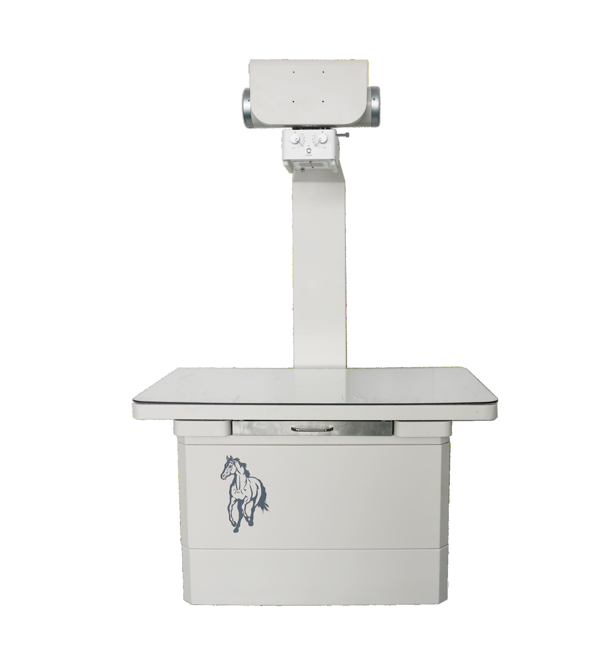
The combined unit is the workhorse answer for a single-room small animal clinic: a table, a column-mounted generator, and castors under the base so the unit can be repositioned or pushed aside when the room needs to serve another purpose. The detail that matters most for veterinary work is the cassette tray that pulls out under the table top.
That tray is not a minor convenience — it is the answer to the non-cooperating patient. A restrained dog on the table does not hold position while a technician fumbles a panel underneath; the tray lets the detector slide into a fixed, known position in seconds, so the restraint window and the exposure window line up. The same tray accepts film cassettes, CR plates, or a digital panel, which means a practice can start with the imaging medium it already owns and move to digital without replacing the table.
Who it fits: first-time buyers, single-location clinics with moderate caseloads, and any practice where the radiography space must do double duty. The adjustable column is the other veterinary-specific feature: table-top studies of a large dog and under-table studies through the tray need the generator head at very different heights, and a column that adjusts across that range saves repositioning time on every study.
Configuration 4: the fixed veterinary DR room
At the top of the range is the fixed veterinary DR system: a floating table, a high-power-class tube on a column, and a touchscreen console that drives the workflow. This is the configuration for practices where radiography is not an occasional event but a daily production line — busy referral hospitals, dental-heavy practices, orthopedic cases, oncology staging, and teaching institutions.
Three features earn their keep at this volume:
- The floating table. Repositioning a sedated large dog by hand is slow, hard on staff backs, and disturbs the positioning you just set. A tabletop that glides in both directions lets the team slide the patient into position and lock it — faster, gentler, and safer.
- The touchscreen console. When the same team runs dozens of studies a day, technique selection, view protocols, and retake handling should live at the table, not on a separate workstation across the room.
- Consistent geometry. A fixed installation means the same source-to-image relationship on every study, which is what makes follow-up comparisons and technique charts actually work.
Who it fits: high-volume practices, specialty and referral hospitals, institutions, and any clinic planning to add dental or advanced imaging that benefits from a stable, dedicated room.
What to check on any configuration: the veterinary-specific details
Whatever the configuration, a handful of details decide day-to-day satisfaction more than the headline numbers on a brochure:
- Table height and patient transfer. The table must accept a large dog lifted by two people, or a patient on a stretcher, without the team stooping dangerously. Check the height against your tallest and heaviest patients, not an average.
- Panel coverage versus your largest patient. A panel that comfortably covers a cat can leave a deep-chested dog requiring multiple exposures and stitching. Match panel size to the biggest patient you actually see, not the species label on the door.
- Duty cycle for repeat series. Veterinary studies frequently involve multiple views in quick succession on a sedated patient — the window closes when the sedation lightens. A generator and tube that need long cooling pauses between exposures quietly sabotage exactly those cases.
- Cleaning and disinfection. Fur, saliva, and the occasional accident are part of the job. Surfaces that wipe down easily, and electronics protected from splashes, age far better in a veterinary room than in a human one.
- The restraint hardware picture. Troughs, sandbags, Velcro straps, positioning foam — plan where these live and how they reach the table. A machine without a restraint workflow around it is half a machine in this field.
The restrainer is the dose problem: a word on radiation safety
This deserves its own section because it is the single biggest difference between veterinary and human imaging practice. When an animal cannot be positioned by equipment alone, a person stands beside the table during exposure. That person — often the most experienced technician, precisely because gentle handling is a skill — is the individual most exposed to scatter in the whole practice.
Responsible answers combine equipment and policy:
- Distance first. Scatter falls off rapidly with distance; every step the restrainer can take back from the table, and every tool that removes their hands from the beam — troughs, straps, foam wedges — buys real protection.
- Protection worn, not owned. Aprons, gloves, and thyroid shields only work when they are worn for every exposure, and gloves are inspected for cracks. Audit this; do not assume it.
- Sedation policy. Many practices find that a defined sedation protocol for uncooperative patients does more for image quality and staff safety than any equipment upgrade — fewer retakes, fewer hands in the beam, shorter restraint windows.
- Training and monitoring. Positioning drills and personal dosimetry make the safety program real instead of theoretical.
Honest framing: equipment can reduce the need for manual restraint, but no buyer should purchase a veterinary X-ray system expecting the safety problem to solve itself. The practices that do this well treat protection as a standing procedure, not a poster on the wall.
Five questions that pick the configuration
- Where does imaging happen today? If the answer is “wherever the animal is” — farms, stables, clients’ homes — the portable case is the conversation. If it is “one room” — combined unit or fixed room. If it is “several places in one building” — the mobile set.
- How many studies per day? Occasional studies forgive a simple setup; daily volume punishes every improvised step. Volume is what turns a tray, a floating table, and a console from luxuries into payback.
- What is the largest patient you actually image? Not the average — the largest. It sets panel coverage, table surface, and generator class in one answer.
- Who holds the patient, and how often? High restraint frequency argues for sedation-friendly workflows, trays, and floating tables — the configurations that reduce hands-on positioning.
- What does the next three years look like? A clinic planning to add dental, orthopedics, or referral work should buy into a configuration that grows — digital-ready trays, upgrade paths, room space — rather than the cheapest answer to this year’s caseload.
Where this guide stops being enough
Three honest boundaries. First, equine and large-animal work is a discipline of its own — the portable generator travels to the horse, but standing-limb techniques, high-output requirements, and field safety deserve specialist attention beyond this article. Second, radiography is one imaging tool among several: for soft-tissue questions, ultrasonography answers things radiography cannot, and most well-run practices own both. Third, the detector is half of every DR decision — wired or wireless, size, and workflow fit — and that choice is covered separately in a field-tested decision framework for wired or wireless detectors in a veterinary clinic.
Frequently asked questions
Can a human portable X-ray unit be used in a veterinary clinic?
Physically, imaging is imaging — but veterinary work imposes demands a human-oriented configuration may not anticipate: no cooperation from the patient, restraint workflow, wide size range, and frequent series exposure. Equipment designed or configured for veterinary use anticipates these from the start, which is why the veterinary line exists as its own category rather than a relabeled human one.
What is the difference between the combined unit and a fixed DR room?
The combined unit is a table, generator, and tray on castors — it can move, serve a multi-purpose room, and start with modest imaging media. A fixed DR room is an installed system with a floating table and console built for daily volume. The practical divider is caseload: below a certain daily study count the combined unit is the better value; above it, the fixed system’s speed pays for itself.
Do I need a flat panel detector, or can I start with CR?
You can start with CR — the tray on the combined unit accepts cassettes, CR plates, and digital panels, so the table does not force the imaging medium. The upgrade to digital is mostly a detector decision, and that decision has its own framework (linked above): workflow volume, retake rates, and whether wireless flexibility matters in your rooms.
Is a mobile X-ray set useful in a small clinic?
Usually not the first purchase. Mobile sets earn their place when imaging must reach several locations — wards, isolation, multiple rooms. A single-room clinic is generally better served by one well-chosen table setup, and the budget difference buys restraint hardware and staff training that improve every study.
How do I know which configuration fits before I buy?
Write down the five answers: where imaging happens, studies per day, largest patient, restraint frequency, and the three-year plan. Send them to the supplier and ask them to recommend a configuration against those answers — a serious manufacturer will engage with the caseload question, not just quote a box. The veterinary system range covers all four configurations precisely so that the recommendation can follow the caseload instead of the other way around.
Talk to us about your animal mix
Tell us the species you see, the largest patient on your table, and how many studies your team runs in a week — we will help you match a configuration to that reality rather than to a brochure. Reach the team at the contact page, by email at admin@newheek.cn, by phone or WhatsApp at +86 19062611512, or through an appointment request.