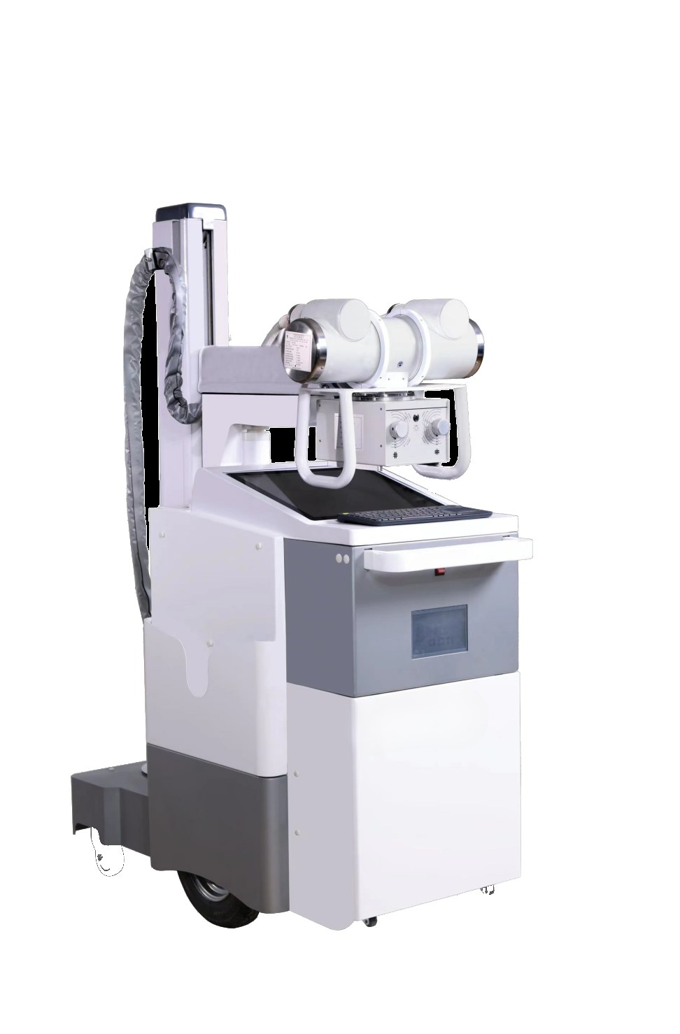
Quick answer: a mobile X-ray machine is a complete radiography system built on wheels — tube, generator, detector, and controls all in one unit you can push down a corridor and park next to a hospital bed. Instead of bringing the patient to the radiology department, the department comes to the patient. The trade-offs are real (tight spaces, battery discipline, infection control), but for ICU patients, isolation wards, nursing homes, and any patient who cannot safely be moved, it is often the only exam that can happen at all.
Most people never think about this, but a standard X-ray room has one quiet assumption baked into it: the patient will come to the machine. There is a table, a wall stand, a generator bolted behind the wall. Everything waits in place.
Now take a patient in intensive care. Two lines in the throat, a pump on each side of the bed, a nurse for every two patients. Moving that patient to the basement radiology suite means disconnecting equipment, coordinating three staff members, and taking a fragile person on the least therapeutic elevator ride of their life.
So the machine goes to them. That is the entire idea behind mobile radiography, and it is worth understanding how it actually works before you buy one — because the workflow is more different from a fixed room than most buyers expect.
Why the Machine Travels
The classic settings for mobile X-ray all have one thing in common: the trip to radiology is either dangerous, impossible, or pointless.
- Intensive care. Chest X-rays on ventilated patients are one of the most frequent imaging exams in any hospital, and most of those patients cannot travel. The film comes to the bed, usually once a day or whenever the care team needs to check lines and lungs.
- Isolation and infectious wards. Wheeling a contagious patient through public corridors is a risk nobody wants. A machine that can be disinfected between rooms is the safer visitor.
- Operating theatres. Surgeons need to confirm hardware position or bone alignment while the patient is still on the table and still asleep.
- Emergency and trauma bays. Some patients are simply too unstable to move until they are stabilised — and stabilising requires images first.
- Nursing homes, wards in older buildings, and field clinics. Anywhere the population cannot easily come to the machine, the economics flip: one machine serves many beds.
Notice what is not on that list: routine outpatient imaging. If a patient can walk to the radiology department, they should — a fixed room gives better consistency, faster throughput, and lower cost per exam. Mobile units earn their keep in the specific situations above, and a buyer who is clear about that ends up much happier with the purchase.
What Actually Happens During a Bedside Exam
This is where first-time buyers get surprised. A mobile X-ray exam is not “a normal X-ray, just wheeled in.” The whole choreography changes.
1. Getting there is part of the job
The machine rides elevators, crosses thresholds, and turns in corridors that were never designed for it. This sounds trivial until you measure a hospital corridor. Wheel diameter, turning radius, and overall footprint are not brochure decoration — they decide whether the machine can reach the fourth floor of a 1980s building at all. Ask to see the actual dimensions, then walk your own route and measure the tightest corner between the elevator and the worst ward.
2. Power: outlet or battery
Older mobile units plug into wall outlets, and finding a free socket next to a bed crowded with infusion pumps and monitors is its own small adventure. Newer designs run on batteries, which changes the job completely: the machine arrives ready, stays ready for a shift, and charges between rounds. The question to ask any supplier is not “does it have a battery” but “how many exposures on a full charge, and what happens to the day’s schedule when it runs flat at bed number nine.” Battery discipline is now a real operational skill on busy wards.
3. The detector goes behind the patient
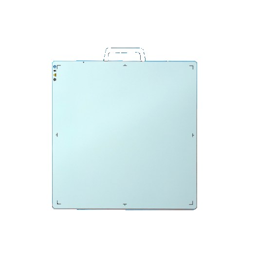
In a fixed room, the detector sits in a table or wall stand. At the bedside, a technologist slides a flat panel detector behind the patient — between the patient and the mattress, with a gown and a pillow on top for comfort. This is one of the gentlest parts of the exam and one of the most physically demanding: the patient may be barely able to move, and the technologist is lifting and positioning the detector by hand, over and over.
Detectors built for bedside work are lighter, have grab handles, and survive being handled all day. If the mobile machine you are considering comes with a detector, handle it yourself. Lift it. Imagine doing that thirty times a shift.
4. Collimation matters even more in a crowded room
A fixed radiography room is built with shielding in the walls. A ward is not. In a shared ward, the exposure field has to be confined tightly to the area of interest, because the people standing three metres away are not patients who consented to anything — they are the patient in the next bed and a nurse mid-task. Good technique here is unglamorous and decisive: tight collimation, sensible distance, and clear communication before every exposure. The collimator on the tube head is doing quiet, constant work every single time.
5. The image travels back like everything else
A mobile exam that ends with “we’ll bring the images down later” is a workflow from twenty years ago. Modern units send the image straight to the hospital’s PACS over Wi-Fi, so the ICU physician sees the result at the same workstation as always. Before buying, test this specifically: how does the unit connect, what happens in a dead zone of the building, and does the image land in the same workflow as exams from the fixed rooms? A mobile machine that feeds the same image chain as the rest of radiology is worth far more than a slightly cheaper one that lives on its own screen.
The Things Nobody Puts in the Brochure
After enough conversations with radiographers who run mobile rounds daily, the same honest observations come up:
- Space is the real constraint. The machine does not need a room; it needs a parking spot, a charging corner, and a path. Hospitals that figure out where the unit lives between rounds get far more use from it than hospitals that treat it as “the machine in the corridor.”
- Cleaning between patients is part of the exam. In isolation settings, the machine must be wiped down between wards. Smooth surfaces, sealed controls, and no fabric crevices are infection-control features, not styling choices.
- Technique flexibility matters at the bedside. Patients at the bedside cannot stand, cannot always lie flat, and cannot always cooperate. The exposure settings and tube positioning need enough range to handle a chest X-ray on a seated, restless patient at a moment’s notice. This is where a purpose-built mobile unit pulls ahead of improvising with lighter portable gear — more on that difference below.
- The schedule is the product. A mobile unit’s value is measured in exams delivered per shift with the battery alive and the technologist not exhausted. Reliability and turnaround are the specs that matter most, even though they never appear on a spec sheet.
Mobile vs Portable vs Handheld: A Quick Map
These three words get used interchangeably in listings, and mixing them up is the most common buying mistake in this category. The short version:
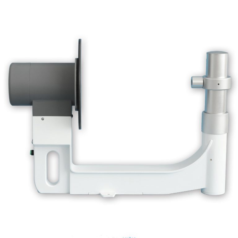
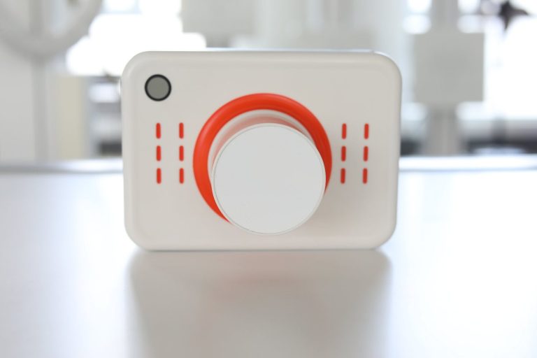
- Handheld units are small devices you literally hold — mainly dental, plus a few low-power applications. They fit in a case, not on a cart.
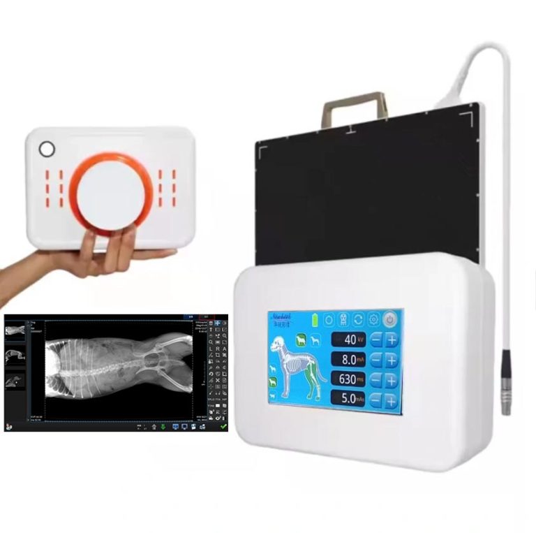
- Portable units are compact systems light enough for one person to carry and set up — popular with clinics, home-visit services, veterinary practices, and outreach programs. They trade some power and automation for mobility.
- Mobile units are the full-size wheeled systems described in this article: hospital-grade output, professional detectors, PACS integration, built for daily rounds across many beds.
We have broken down the full differences between the three devices sold under the “handheld X-ray” name separately — if you are still deciding which class of machine you actually need, start there. This article assumes you already know it is the wheeled mobile class, or you are seriously considering it.
Who a Mobile X-Ray Machine Is Actually For
Honest fit assessment, from the field:
- Hospitals with ICU, surgery, or large long-term-care populations: the clearest yes. The unit pays for itself in avoided patient transports and faster clinical decisions.
- Nursing homes and rehabilitation centres: increasingly common buyers. Residents with dementia or limited mobility often cannot be transported for a simple chest film; one mobile unit serving several facilities is a working business model.
- District clinics and outreach programs: usually better served by a portable unit that fits in a vehicle — unless the clinic serves an immobile population on site.
- Buyers who mostly want “a cheaper fixed room”: usually a mismatch. A mobile unit is not a budget radiology room; it is a different tool with its own discipline. If your volume is walk-in outpatients, build or upgrade the fixed room first.
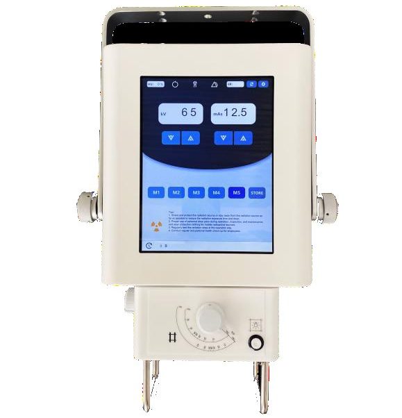
Within the mobile class itself, machines vary widely — from compact units suited to nursing homes up to hospital-grade systems with full detector workflows, like the mobile X-ray machine in our product range. The right comparison is not “which machine is strongest on paper” but “which one matches the corridor widths, the daily exam volume, and the image workflow of your actual facility.”
Six Questions to Ask Before You Request a Quote
- Route first, specs second: what are the machine’s exact dimensions, wheelbase, and turning radius — and can it reach the worst ward in your building?
- Battery reality: how many exposures per full charge, how long to recharge, and can it run on AC while parked when the battery runs low?
- Detector handling: is the detector wireless, how much does it weigh, and is it built for repeated bedside positioning?
- Image workflow: does the unit send images directly to PACS or a workstation, over what connection, and with what happens-if-it-drops plan?
- Infection control: are surfaces sealed and wipeable, and are there documented cleaning procedures between wards?
- Compliance and documentation: what registration or regulatory documents does the unit come with for your country — ask for the list in writing before you commit.
A supplier who answers these six plainly — including the uncomfortable ones — is a supplier worth shortlisting. One that only sends a spec sheet and a price is selling you a box, not a working mobile service.
Frequently Asked Questions
Is a bedside X-ray as good as one taken in the radiology room?
For the exams mobile units are used for — mainly chests, lines and tubes, and orthopaedic follow-ups — yes, clinically sufficient. Image quality can be slightly more variable because positioning and patient condition are less controlled, but the alternative for these patients is no image at all. Radiographers compensate with technique, and the diagnostic value comes from answering the clinical question, not from winning a beauty contest.
How long does a mobile X-ray exam take at the bedside?
The exposure itself takes seconds. The full visit — transport, positioning the detector, explaining to the patient, exposing, confirming the image — typically runs a few minutes per bed, longer if the patient needs careful handling. The real planning variable is travel time between wards and elevator waits, which is why round scheduling matters more than exam speed.
Do staff and nearby patients need protection during a bedside exposure?
The same radiation principles apply as anywhere: keep the field tight, keep distance, and follow your facility’s shielding procedures for staff and neighbours. The exposure field is collimated to the area of interest, and trained technologists manage the rest of the room before every exposure. Your local radiation safety officer sets the specific ward procedures — a good supplier supports that conversation with documentation rather than dismissing it.
Can one mobile unit serve several buildings or facilities?
Yes, and it is a common model for nursing home groups: the unit travels a route, and each site books a window. The requirements are a vehicle or covered path between sites, a charging spot at each stop, and predictable scheduling. Battery capacity becomes the route-planning constraint — count exposures per site and check them against a full charge.
What maintenance does a mobile X-ray machine need?
Plan on periodic calibration and inspection per your country’s requirements, battery health monitoring over the years, and detector care similar to any digital radiography system. Ask the supplier what service support looks like in your region before buying — response time for a down unit matters more for a mobile service than for a fixed room, because there is no backup machine to walk the patient into.
Talk to a Supplier Who Has Pushed One Down a Corridor
Choosing a mobile X-ray machine is less about one spec and more about the fit between the machine and your building, your patients, and your daily rhythm. If you send us your corridor widths, the floors the unit must reach, and roughly how many exams per day you expect, we will come back with a concrete configuration and the documentation list for your market — no template quotes.
Email: admin@newheek.cn
Phone / WhatsApp: +86 19062611512