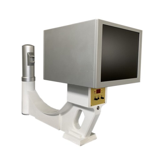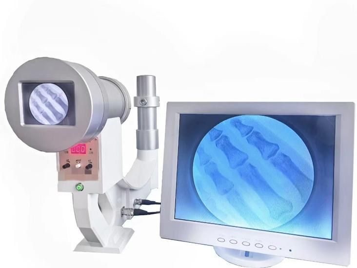
Quick answer: The kilowatt figure on a portable X-ray unit is the ceiling of the kV × mA product — it is not a penetration grade. Between a 5 kW and a 5.6 kW unit, the real difference you will live with is not the 0.6 kW; it is the mAs range, the exposure-time range, weight, and voltage flexibility. In the three portable configurations on this site, the highest-output option tops out at 0.1–320 mAs against 0.1–100 mAs on the lightest unit — that difference decides whether you can image a thick abdomen or a large dog on the first exposure, far more than the nameplate power does. This article reads the published specifications line by line so you can match a configuration to your workload before you compare quotes.
Generator power is the first number buyers see and the last one they understand. Sales pages lead with it, quotes rank by it, and yet two units with nearly identical power ratings can behave very differently on a bariatric chest or a canine hip. The confusion is understandable: power ratings compress four separate realities — voltage ceiling, current ceiling, exposure-time range, and thermal headroom — into one flattering number. This article unpacks what each rating actually buys, using the published specifications of three portable configurations we manufacture, so the next “5 kW vs 5.6 kW vs 8 kW” comparison you run is decided by workload, not by marketing.
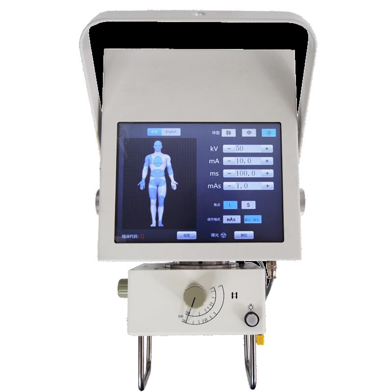
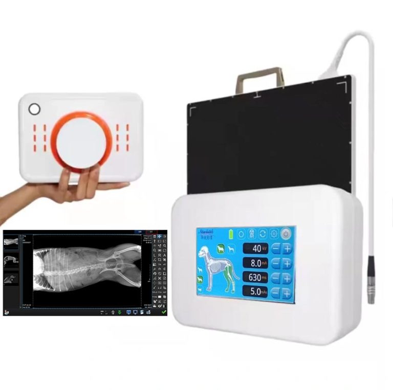
A portable unit’s touchscreen console: kV, mA, ms, and mAs are the four numbers that actually shape the exposure — the nameplate kW only bounds their combination.
What the “kW” Figure Actually Measures
A generator’s power rating is, at its core, the product of tube voltage and tube current at the point of exposure: roughly, kilovolts × milliamperes defines whether the machine can deliver its rated output simultaneously. A “5 kW” rating with a 40–125 kV range and a 10–100 mA range means the generator can push meaningful current at the top of the voltage range — that combination is what the rating certifies.
Three practical consequences follow, and they matter more than the headline number:
- The kV ceiling is a separate decision. Two of the three configurations on this site reach 125 kV; one tops out at 120 kV. Whether your thickest patient is imaged comfortably or not is decided by that ceiling, not by whether the nameplate says 5.0 or 5.6 kW.
- Output headroom is expressed in mAs, not kW. The mAs range — the product of current and time you can accumulate in one exposure — is what lets you push through a thick shoulder or a sedated large animal. That range varies by a factor of more than three across same-power units.
- The gap between “rated” and “maximum” output is small. Our published specs list 5 kW (maximum 5.3 kW) and 5 kW (maximum 5.6 kW) on different configurations — the maximum ratings differ by about 6%, which no image ever showed.
None of this means power ratings are fake. It means they are a boundary condition, and the interesting purchasing questions live inside the boundary.
kV Range: The Real Penetration Variable
Kilovoltage is what pushes the beam through tissue. Published specs on the three configurations read as follows (all values from the product pages; confirm against the datasheet of the exact unit before purchase):
- The compact 5 kW unit (product page): 40–125 kV, adjustable in 1 kV steps.
- The 5.6 kW unit with four interface languages (product page): 40–125 kV, 1 kV steps.
- The dual-voltage 5.6 kW unit (product page): 40–120 kV, 1 kV steps.
Read that list carefully: the 5 kW unit and one of the 5.6 kW units share an identical 40–125 kV ceiling. For extremity, chest, pediatric, and small-animal work, all three configurations operate in the same comfortable band. The difference only appears at the top end — a lateral lumbar spine on a heavyset patient, or a large dog’s hip — where those extra 5 kV on the 125-kV units shorten exposure time and reduce motion blur.
The 1 kV step is worth a sentence too. Fine voltage steps let you tune technique charts to your detector and your patient mix instead of rounding to whatever coarse steps an older generator offered. Every configuration here supports it; if you evaluate units from other suppliers, verify it, because coarse steps are a common cost cut.
mAs Range: The Quiet Differentiator
If kV decides whether the beam gets through, mAs decides how much information comes back. Here the three configurations genuinely separate:
- Compact 5 kW unit: 0.1–200 mAs, with an exposure-time range of 1–4,000 ms.
- 5.6 kW four-language unit: 0.1–100 mAs.
- Dual-voltage 5.6 kW unit: 0.1–320 mAs, with exposure times from 10–6,300 ms.
What does a 320-mAs ceiling buy that a 100-mAs ceiling does not? Headroom for the thick and the uncooperative: large patients where you need maximum penetration at adequate detector exposure, veterinary patients whose anatomy does not conform to a human technique chart, and situations where you would rather extend exposure time than raise current. The 10–6,300 ms range on the dual-voltage unit similarly accommodates long, low-current exposures where a short burst would freeze nothing but cost image quality.
The floor matters as much as the ceiling: all three configurations reach down to 0.1 mAs, which is what keeps pediatric and dental-adjacent work clean and low-dose. A wide range at both ends is the sign of a well-specified generator — and it is invisible in the kW number.
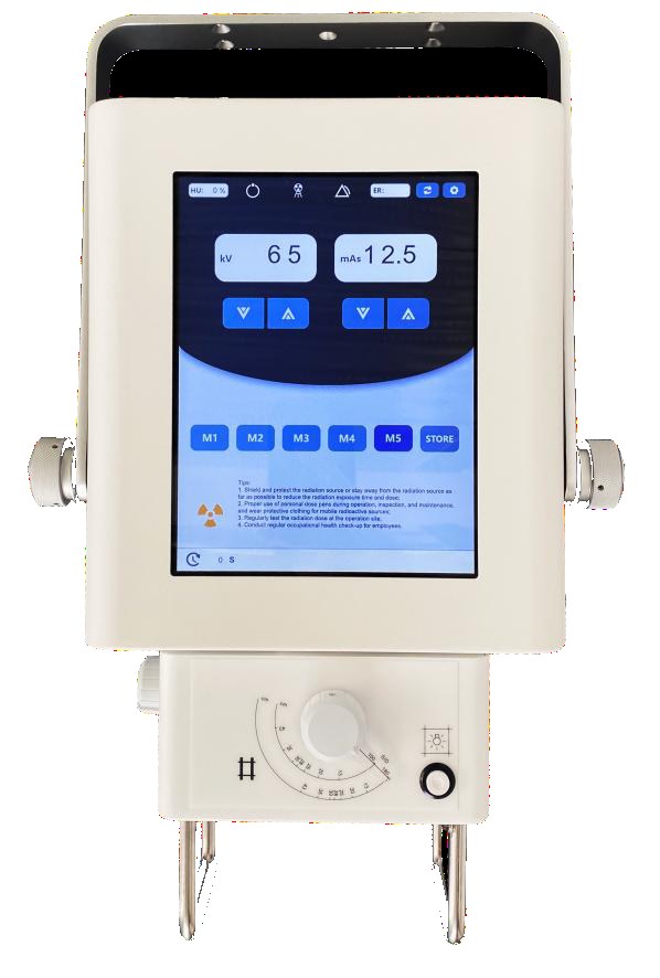
kV and mAs on the console: the two numbers a technologist actually balances on every exposure. Published mAs ceilings differ by more than 3× across same-power portable units.
Weight, Voltage, and the Workflow Reality
Portable means portable. The compact 5 kW unit weighs 15 kg with dimensions of 265 × 240 × 210 mm; the two 5.6 kW configurations weigh 17.5 kg. Two and a half kilograms sounds trivial until it is being carried up a clinic staircase twice a day — for bedside and field work, the lighter unit earns its keep despite the smaller mAs range.
Voltage flexibility is the other quiet decider. The dual-voltage 5.6 kW configuration accepts 110 VAC / 220 VAC at 50/60 Hz — the published page also notes 8 kW and 12.5 kW versions on request — while the other two run 220–240 VAC. If your market has unstable or mixed mains power, or you are equipping a vehicle or a field program in a 110 V region, that single specification line can overrule everything else in the comparison.
Smaller comforts add up the same way: the four-language unit ships with Chinese, English, Russian, and Spanish interface options; all three configurations expose hand-switch and remote exposure control and pair with flat panel detectors, wired or wireless, for digital radiography.
So When Is 8 kW the Right Answer?
An 8 kW (or 12.5 kW) generator earns its cost in three situations:
- Consistently thick anatomy. Bariatric imaging, large-breed veterinary work, and equine limbs where every 5-kW exposure already sits at the top of the technique chart.
- High patient throughput on thick views. A busy clinic shooting abdomens all day benefits from shorter exposure times at equivalent mAs — less motion blur, fewer repeats.
- Future headroom. If detector upgrades or new service lines are planned, buying generator headroom once is cheaper than replacing a unit later.
For extremity, chest, pediatric, dental-adjacent, and small-animal work, a well-chosen 5 kW configuration does the job at lower cost and lower weight. The honest answer for most buyers: your workload decides, and the technique chart you already run tells you which side of the line you are on.
Side-by-Side: The Three Configurations at a Glance
| Specification | Compact 5 kW | 5.6 kW (four languages) | 5.6 kW (dual voltage) |
|---|---|---|---|
| kV range | 40–125 kV, 1 kV step | 40–125 kV, 1 kV step | 40–120 kV, 1 kV step |
| mAs range | 0.1–200 | 0.1–100 | 0.1–320 |
| Exposure time | 1–4,000 ms | — (see product page) | 10–6,300 ms |
| Weight | 15 kg | 17.5 kg | 17.5 kg |
| Mains voltage | 220–240 VAC | 220–240 VAC | 110/220 VAC, 50/60 Hz |
| Interface languages | CN / EN / ES | CN / EN / RU / ES | — (see product page) |
| Focal spots | 0.6 / 1.8 mm | 0.6 / 1.8 mm | — (see product page) |
| Upgrade path | — | — | 8 kW / 12.5 kW versions on request |
All values are quoted from the published product pages linked above; treat them as the starting point for a conversation, and confirm the datasheet of the exact configuration before you order.
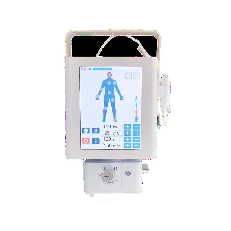
Exposure parameters on the console. The published ranges behind these controls — not the nameplate kW — are what your technique chart will live on.
A Note on Scope: Detector and Mounting Questions Live Elsewhere
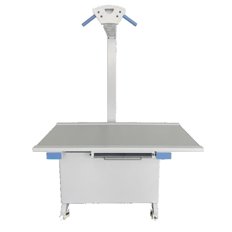
This article deliberately stays at the generator-and-workflow level. If you are comparing image quality at the sensor level — pixel pitch, scintillator type, wireless vs wired links — those belong to the detector itself, and the engineering detail is covered in depth on our detector-focused site. Likewise, stands, wall mounts, and room planning follow their own logic. What a portable unit contributes to that chain is a stable, repeatable exposure — everything in this article serves that goal.
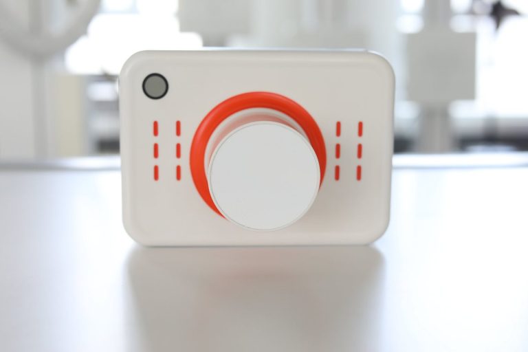
If your interest is the genuinely handheld category — dental units that operate one-handed at chairside — that is a different device class with different physics and different regulations; our overview of the handheld dental X-ray machine draws that boundary in detail.
FAQ
Is a 5.6 kW unit automatically better than a 5 kW unit?
No. The 0.6 kW difference is a generator ceiling, not an image-quality grade. Compare kV range, mAs range, weight, and voltage options against your workload: the compact 5 kW unit in this comparison actually reaches a higher mAs ceiling (200 vs 100) than the lighter-spec 5.6 kW configuration.
What matters more for thick patients: kV or mAs?
They work together, but kV decides whether the beam penetrates at all; mAs decides how much image information returns. For consistently thick anatomy, prioritize the 125 kV ceiling and the widest mAs range you can find — on this site, the dual-voltage 5.6 kW unit’s 0.1–320 mAs range is built for exactly that case.
Do I need the 8 kW version?
Only if your technique chart already runs at the top of a 5 kW unit — bariatric, large-animal, or high-throughput thick-view workloads. For extremity, chest, pediatric, and small-animal imaging, a 5 kW configuration is usually sufficient, lighter, and more economical. The 8 kW and 12.5 kW versions are available on request on the dual-voltage product line.
Why does mains voltage matter on a portable X-ray unit?
Because portability often means crossing power standards. The dual-voltage configuration accepts 110/220 VAC at 50/60 Hz, which makes it usable in mixed-standard regions, vehicles, and field programs without external conversion equipment. Units fixed to 220–240 VAC need a stable mains supply of that standard.
Can these portable units be used for digital radiography?
Yes. All three configurations pair with flat panel detectors, wired or wireless, per their published product pages, and expose hand-switch and remote exposure control. The detector determines image quality at the sensor level; the generator determines whether the exposure is repeatable at the dose you planned.
Talk to Us
Tell us your patient mix and thickest routine view, and we will point you at the configuration whose published range covers it — or configure one that does:
Email: admin@newheek.cn
Phone/WhatsApp: +86 19062611512 (WhatsApp chat)
Or leave a message via our contact page.